Tag
16 april 2026

How MERLN


creates biomedical magic

At the research institute MERLN, scientists are working magic with regenerative medicine. They’re developing groundbreaking treatments that help restore proper function in diseased cells, tissues, and organs. Meet the researchers behind MERLN and marvel at some of their most impressive projects.

MERLN’s headquarters at Universiteitssingel 40 in Maastricht.

What if a complex bone fracture could heal itself? What would it be like if animal testing were no longer necessary, because we could create hearts and other organs in the lab? And what if we not only understood why we age, but could actually stop the process? It may sound like science fiction, but thanks to MERLN, breakthroughs like these are closer than we think.

The full name of the research institute is ‘MERLN Institute for Technology-Inspired Regenerative Medicine’. Regenerative medicine is a new therapy approach that helps restore diseased cells, tissues, and organs to health or replace them. Examples include heart problems, kidney diseases, or broken bones and ruptured tendons. The approach uses the body’s own natural healing capacity.


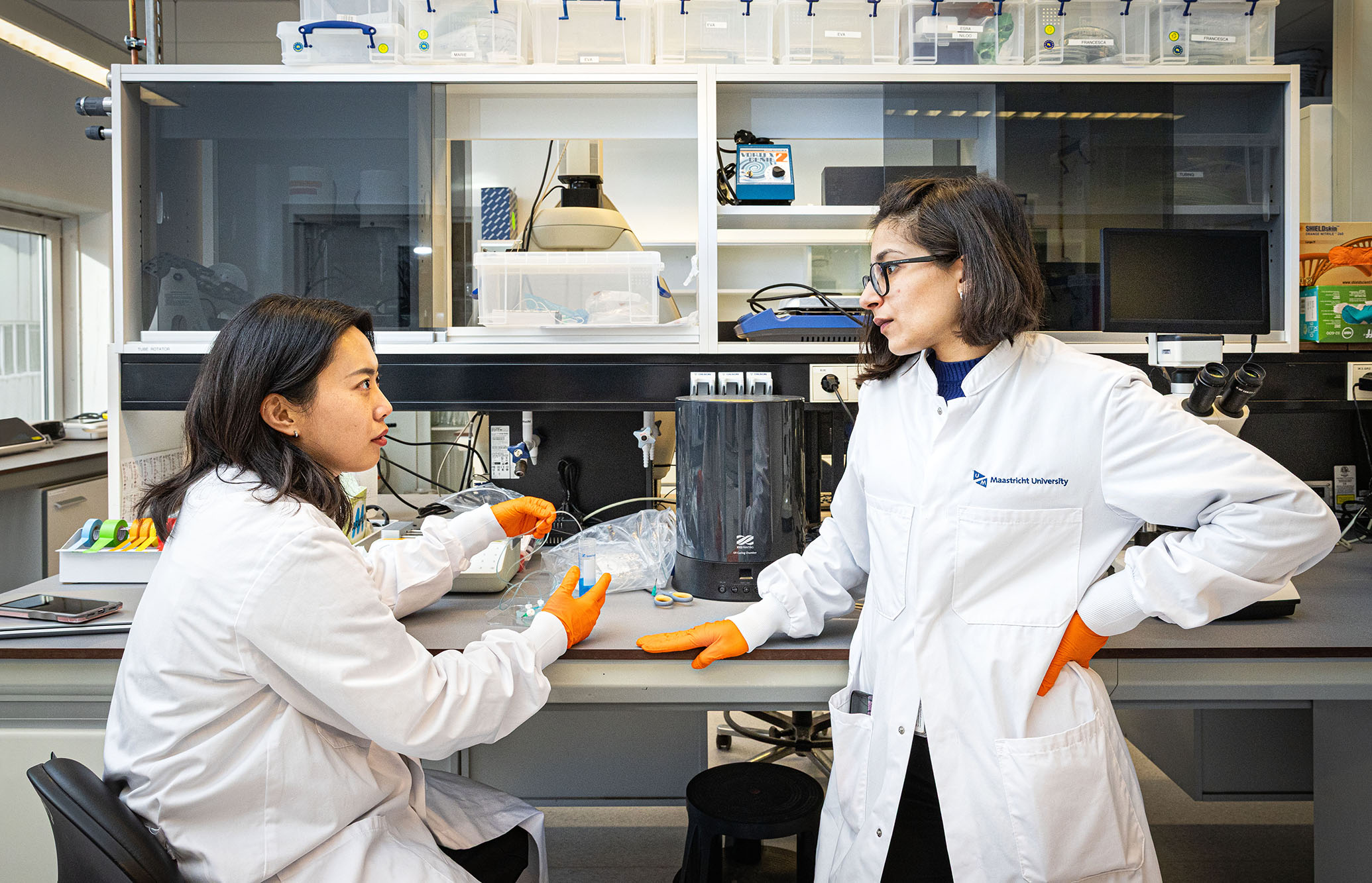
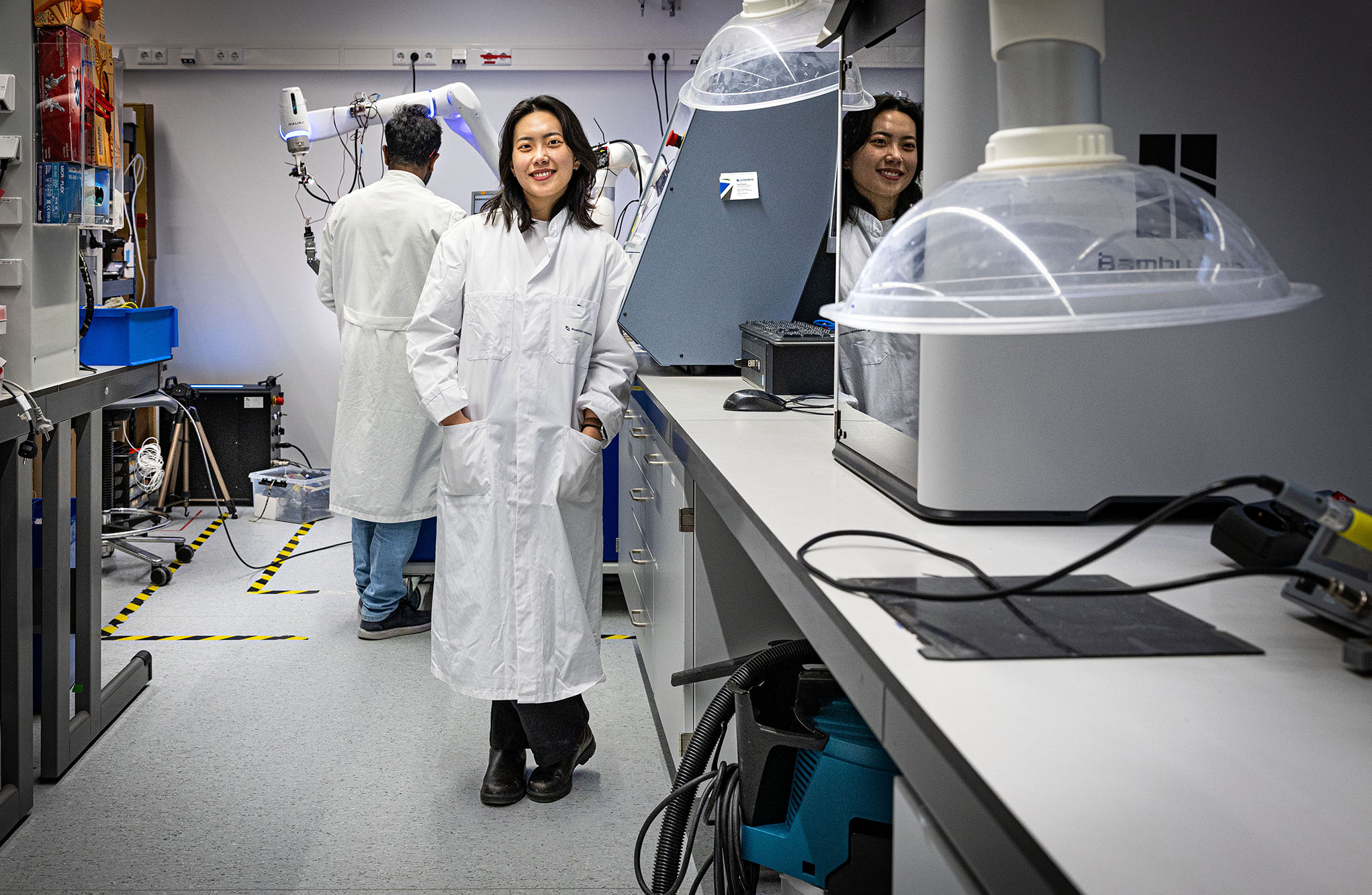
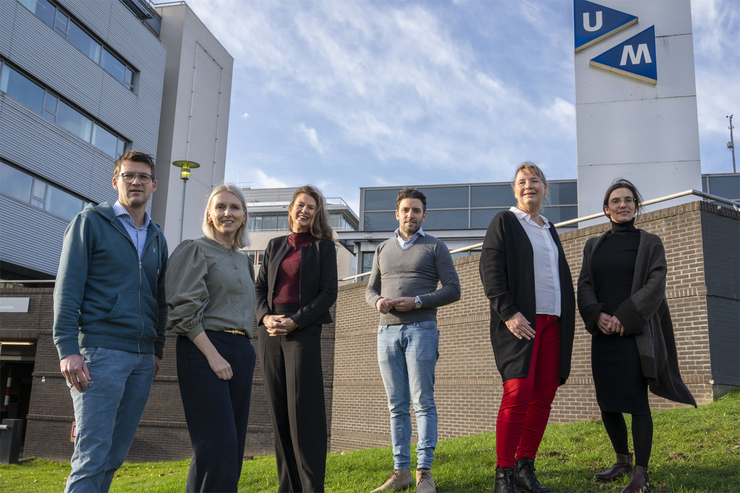
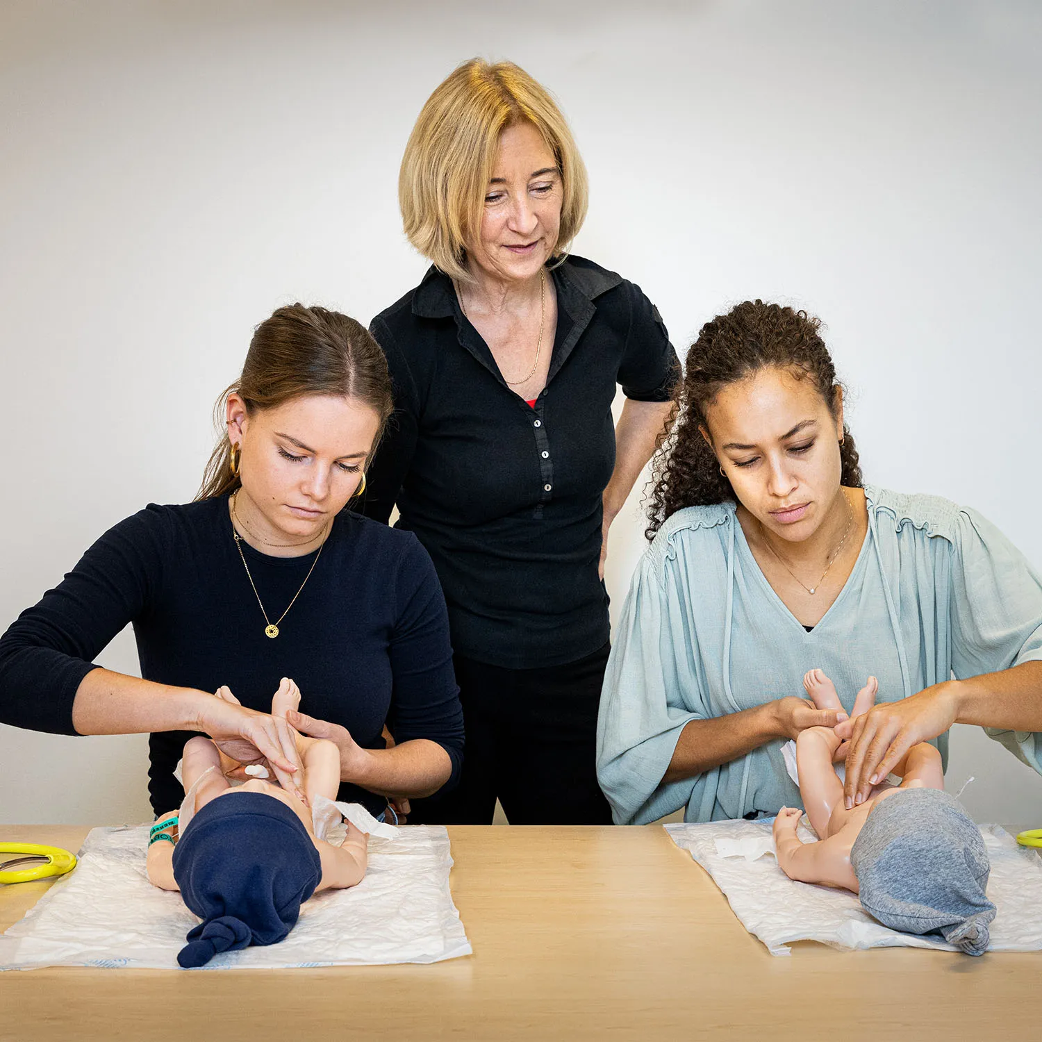
At MERLN, researchers from a wide range of backgrounds work together on new techniques – from chemists to biomedical engineers. MERLN is also very international: the researchers come from more than thirty different countries!

That diversity could create a sense of distance, but at MERLN the opposite is true. There is a close-knit, vibrant community where colleagues easily find common ground.


Every Friday, for example, researchers share their latest insights during lunchtime presentations. This keeps everyone engaged with each other’s work.


Bones and tendons that heal themselves

What if damaged tendons and bones could repair themselves? No matter how severe the damage? That is what Carlos Peniche is working on. His research aims to understand the molecular mechanisms responsible for the healing of musculoskeletal tissues, to make them heal better and faster.

He evaluates the use of structures made of special synthetic or biological materials. For example, imitating the structure of a tendon to improve the treatment of injuries. He places stem cells inside these structures with specific molecules. These molecules signal the cells to grow into new bone, tendon, or connective tissue.


Carlos Peniche

Bones and tendons that heal themselves

One of Peniche's students is working on a structure made of bone material.

The ultimate goal is to implant these structures, containing biomolecules and living cells, into the body – precisely at the site of the damage. There, the cells set to work to create new tissue.

When it comes to bones, Peniche collaborates with other researchers using not only 3D-printed structures, but also actual bone material. Using bone increases the likelihood of treatment success.

If someone has a very complex bone fracture, doctors sometimes can’t repair it. A broken arm or leg must then be amputated. Thanks to this kind of research, this may soon no longer be necessary.


From left to right: a 3D-printed plastic structure for living cells, one  made of bone material, and artificial entheses (fibers that attach tendons to bone).


Cross-section of a rat’s bone attached to a tendon by entheses.

Peniche’s research work could also improve existing treatments. “No two accidents are the same”, says Peniche. “Every injury should receive a custom-made approach. That’s what we’re working on.


A device that Peniche uses to 'read' cells. This allows him to see whether tissues are actually recovering.


Living plastic

To conduct effective research at MERLN, scientists need materials that are as close as possible to real human tissue. That is why they often work with hydrogels: polymers that contain a lot of water, just like the tissues of the human body. Anna Pierrard is investigating how she can make these hydrogels truly lifelike.

Anna Pierrard

Living plastic


“In my research group, we are trying to develop dynamic hydrogels”, explains Pierrard. “These are gels whose properties can change over time. Just like in the human body, where tissues are constantly in motion and adapting.”

Pierrard is carrying a container of liquid nitrogen, which she uses in the lab to cool solutions or liquids quickly.

These hydrogels are colourless, but Pierrard adds a nice touch of colour to them for photos in papers. This makes them easier to see.


Researchers can make the hydrogels softer or stiffer by changing their properties to match different target tissues. After all, cartilage feels very different from a kidney. The ultimate aim is to place hydrogels inside the body to replace damaged or lost tissue. For example, cartilage in the knees, which doesn’t fully regenerate itself.


A closer look at fertilisation

Jaehyeon Kim is aiming to help women struggling to conceive. Through her research, it could be possible to enable better care in the future. She focuses on the fallopian tubes, a part of the female body that remains under-researched.

“A lot of research has always been done on the uterus, because that’s where the baby grows,” says Kim. “But fertilisation takes place in the fallopian tube. That’s where the sperm and the egg meet. And that’s precisely where complications often arise, for example due to infectious diseases such as STIs.”

Jaehyeon Kim

A closer look at fertilisation

Kim is checking that the various fluids in her fallopian tube model aren’t leaking accidentally.

Kim is currently studying the isthmus, which is the first section of the fallopian tube, closest to the uterus. To do this, she is developing a model of the fallopian tube made from hydrogel. Kim is using this to investigate how STIs affect the tissue. These insights could lead to new and better treatments.

Kim uses a CNC machine to make a plastic mould for her models.

Kim hopes these results might one day help some women avoid IVF treatment. Kim: “IVF can be a good solution, but the success rate is relatively low, it is expensive and can be very emotionally draining. We therefore hope to identify the cause of certain fertility problems so that we can treat them directly.”


A mould for applying patterns to hydrogel, so that its structure resembles the inside of a fallopian tube.


The mystery of the origins of life

Research on human embryonic development is important. Scientists want to know, for example, how an organism develops in a healthy and diseased context.

However, you cannot simply carry out such research using real human embryos, as this raises ethical questions. That is why Anna Peeters and Leila Ashtar at MERLN are working on alternative methods. They are creating simplified models of various embryonic stages from stem cells in the lab, without the use of sperm cells and eggs.

Moreover, Peeters and Ashtar study the effects of chemicals, pollutants or medicinal drugs on early embryonic development. By gathering all this information, they also aim to improve fertility treatments such as IVF in the near future.

Leila Ashtar and Anna Peeters

The mystery of the origins of life

The sheets of plastic that Peeters and Ashtar use are only 0.05 millimetres thick.

Their research group produces customised cell culture tools, including thin polymer sheets with grooves smaller than a human hair. They place a defined number of stem cells into these grooves to form small clusters.

Together with a specific cocktail of chemicals, these clusters of stem cells form structures that are very similar to structures found during human embryonic development. For example, human blastocysts around five to six days after fertilisation.


“These are some of the techniques that allow us to study specific aspects of early development of a human embryo for the first time without using actual embryos”, explains Ashtar. “This opens the door to more extensive research.”

Ashtar holds the master mould for the creation of the cell platforms.


Master mould for the cell platforms.


Floating heart

Ezgi Çevik is developing a truly unique 3D printer. It will soon enable her to print heart tissue. But this is no simple process. To bring heart cells together into a single complex tissue unit, she uses a combination of magnets and sound waves.

Ezgi Çevik

Floating heart


Çevik’s printer is designed to help MERLN assemble organoids. These are miniature organ models made from multiple stem cells used to simulate diseases and test treatments. “At the moment, I’m focusing on the heart”, she says. “But ultimately, my device will be able to print all the body’s tissues.”

Çevik demonstrates one of her devices that makes cells float.

Çevik’s 3D printer uses two techniques to bring the cells together: magnetic and acoustic levitation. “In magnetic levitation, I place the organoids in a chamber between two ring magnets with the same poles facing each other”, explains Çevik. “This causes them to float towards the centre and come together there.”

Acoustic levitation of a small polystyrene ball.

The chambers that Çevik uses for magnetic levitation.  

Using acoustic levitation, she brings the cells together with sound waves. “When two sound waves with the same properties travel towards each other, you can make particles hover between the waves”, she says. “This allows you to capture them together and even move them.”


Time travel in the lab

A baby’s heart works differently from that of a 65-year-old. Cells change as we age. Because heart problems often arise later in life, it is important to research adult heart tissue as well. However, lab-grown tissue must age naturally, a process that takes decades.

That is why Alix Lemaître is developing methods to create adult heart tissue directly. Her colleague João da Silva Ribeiro is taking it a step further: he is investigating how they can develop tissue that resembles that of elderly people.

João da Silva Ribeiro and Alix Lemaître

Time travel in the lab


With these ageing heart models, researchers can test medicines and treatments more effectively. That is why Da Silva Ribeiro is focusing on unravelling the ageing process. Why do we grow older? What causes it? And how can you replicate that process in the lab?

Lemaître is studying a cardiac organoid: stem cells that come together to form a beating mini-heart.


“We actually know surprisingly little about that”, he says. “If we understand better how ageing works, we can also find ways to slow it down or stop it. That could have a significant impact on all age-related diseases. And who knows? Perhaps one day we might even be able to reverse the process.”


An incubator with spinning flasks containing cells. Because the cells are spinning around, they can grow more quickly.


A researcher gives the cells their daily dose of nutrients.